
This test is conducted between 16 and 32 weeks of pregnancy (recommended after 17 weeks) to check for any chromosomal abnormalities in the baby by sampling amniotic fluid from the uterus. It includes tests for all chromosomes (G-band), including Trisomy 21 (Down syndrome), Trisomy 18, and Trisomy 13, as well as microdeletion tests (such as microarray) for small chromosomal deletions that may cause abnormalities in the fetus.
As the age of the couple increases, the likelihood of chromosomal abnormalities in the fetus also rises, which is why many couples opt for amniocentesis, a fetal diagnostic test. Recently, many couples have been opting for NIPT (Non-Invasive Prenatal Testing); however, NIPT is a screening test to determine the likelihood of chromosomal abnormalities, not a diagnostic test. Even if the result is negative, false negatives are possible, meaning the true status is uncertain. Therefore, if you want a definitive result, we recommend Chorionic Villus Sampling (CVS) during early pregnancy or amniocentesis after the 16th week.
Couples who have undergone PGT (Preimplantation Genetic Testing) are strongly advised to choose amniocentesis over CVS, as the latter uses tissue from the placenta. While the risk of miscarriage is very low (about 0.1%), it is important to be aware of this possibility. For issues other than chromosomal abnormalities, detailed fetal ultrasound examinations are effective.
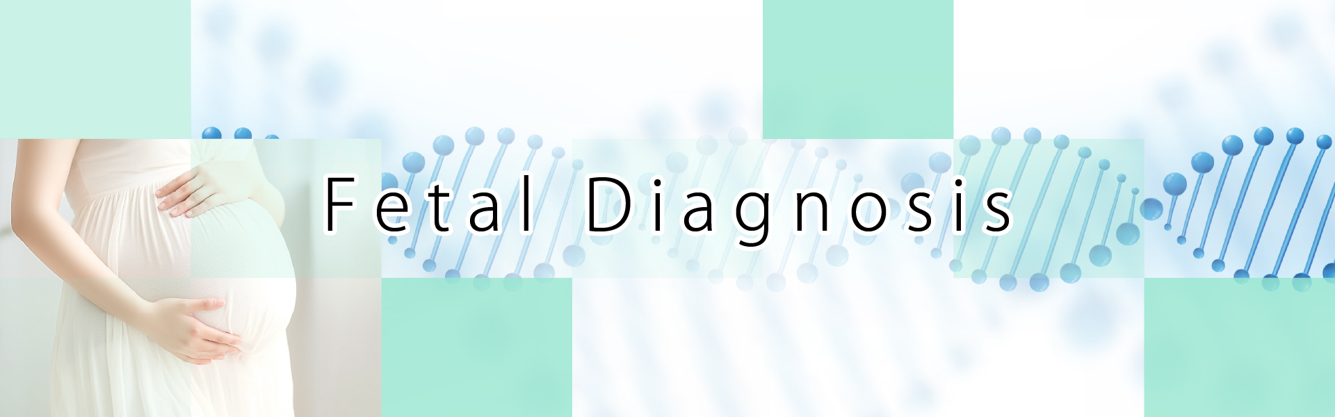
Between 16 and 32 weeks of pregnancy (recommended after 17 weeks), an abdominal ultrasound is used to check the position of the placenta, baby, and umbilical cord. A thin needle is then inserted just below the navel to collect amniotic fluid for testing.
A local anesthetic is applied to the insertion area, so there is minimal pain. Finding the optimal insertion point may take about 10 minutes with ultrasound, but the procedure itself is completed in just 3 to 5 minutes.
The collected amniotic fluid contains fetal cells, which undergo chromosomal analysis (G-band) and microdeletion testing (such as microarray). Results are typically available about two weeks after the test.
After the procedure, you will rest in the clinic for 2 to 3 hours while the baby's condition is monitored before you are discharged. On the day of the test, please avoid bathing and try to rest as much as possible.
▶︎ Chromosomal Analysis (G-Banding)
Standard amniocentesis is performed using a method called G-banding analysis. Fetal cells in the amniotic fluid are cultured to increase their number, and cells in the metaphase stage of division—when chromosome structures are most identifiable—are selected, stained, and analyzed.
This method determines the number of chromosomes 1 through 22, as well as the X and Y sex chromosomes. Since each chromosome is individually examined, large abnormalities such as extra or missing segments and translocations (where chromosome segments are rearranged) can also be detected.
▶︎ Microdeletion Testing (e.g., Microarray)
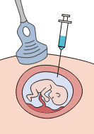
Chromosomes are composed of proteins and DNA (genes).
DNA consists of four types of bases—adenine (A), thymine (T), guanine (G), and cytosine (C)—which form a double-helix structure. The sequence of these bases (base sequence) carries genetic information.
While the number and arrangement of bases are specific to each species, there are regions where the sequence differs from the standard base sequence approximately once every few hundred to a thousand bases.
These single-base differences are called single nucleotide polymorphisms (SNPs), which contribute to individual differences and genetic diversity.
In humans, there are about 10 million SNPs among the 3 billion bases, and several SNPs have been identified as being associated with congenital disorders.
By using a method called "SNP microarray," which analyzes chromosomes at the base sequence level, it is possible to detect subtle chromosomal abnormalities that cannot be identified through G-banding analysis.
Chromosomal microdeletion syndromes vary in symptoms depending on the location of the deletion (Table 1). While some cases may present no symptoms at all, others may involve severe complications.
A well-known example is DiGeorge syndrome, which is associated with organ development abnormalities, congenital heart defects, intellectual disabilities, speech disorders, and immunodeficiency. The prevalence of this syndrome is estimated to be approximately 1 in 4,000 to 5,000 individuals.
|
|
|
|

|
|
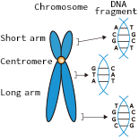
6) Amplifying DNA fragments
↓ 7) Hybridization ↓ 8) Analysis |
9) Can detect even subtle abnormalities |
Table 1. Chromosomal Microdeletion Syndromes
| Name | Chromosomal Deletion Site | Main Symptoms |
|---|---|---|
| Wolf-Hirschhorn Syndrome | Chromosome 4 short arm (4p) | Distinctive facial features, growth retardation, severe intellectual disability, hypotonia, intractable epilepsy, feeding difficulties |
| Williams Syndrome | Chromosome 7 long arm (7q11.23) | Aortic valve stenosis, intellectual disability, 'elfin' facial features, transient hypercalcemia in infancy |
| Prader-Willi Syndrome | Paternal chromosome 15 long arm (15q11) | Hypotonia from birth, obesity, gonadal dysfunction, small hands and feet, intellectual disability |
| Angelman Syndrome |
Maternal chromosome 15 long arm (15q11)
(15q11) |
Seizures, puppet-like ataxia, spontaneous laughter, hand-flapping, severe intellectual disability |
| Miller-Dieker Syndrome | Chromosome 17 short arm (17p13.3) | Lissencephaly, low upward-facing nose, severe developmental delay, seizures, severe intellectual disability |
| DiGeorge Syndrome | Chromosome 22 long arm (22q11.21) | Hypoplasia or absence of the thymus and parathyroids, congenital heart defects, cleft palate, intellectual disability, psychiatric issues |
The test can detect chromosomal abnormalities such as trisomy 21 (Down syndrome), trisomy 18, and trisomy 13, as well as structural chromosomal abnormalities, including small deletions.
Abnormalities in the number of chromosomes tend to increase with maternal age, while small deletions are said to occur randomly, regardless of maternal age.
Additionally, the effects and symptoms on the baby can vary depending on which chromosome has the numerical or structural abnormality. Therefore, it is important to discuss with your partner beforehand what actions to take based on the test results.
Although this is a definitive test, it cannot diagnose all congenital disorders. Other factors, such as contamination of maternal cells, poor culturing of fetal cells from the amniotic fluid, or unexpected reasons, may prevent the test from being completed, and multiple pregnancies can affect the accuracy of the results.
There may also be cases where mosaicism (a mixture of normal and abnormal cells) is detected, or where small chromosomal changes with unclear significance are found.
Our clinic provides genetic counseling and is here to support both of you, so please feel free to consult with us.
| Amniocentesis (including procedure fee) | G-Banding Method | ¥148,000(with tax ¥162,800) |
| +Rapid Method (such as FISH) | +¥20,000(with tax ¥21,000) | |
| +Microdeletion Detection (Microarray) | +¥100,000(with tax ¥110,000) |
※Fees are subject to change without prior notice.
※Results of the test may be delayed. Please understand that this is only an estimate.
※An appointment is required to undergo the test. Please contact us by phone for inquiries.
English Help Desk
TEL:070-1820-0909
Email:english_help@oakclinic-group.com
[Reception time]
Monday-Sunday 09:00-17:00
For urgent inquiries, please contact us by phone.