

Using high-precision ultrasound equipment, which offers greater accuracy than standard prenatal check-up ultrasounds, we conduct detailed assessments of the baby’s condition, including any abnormalities and blood flow. This examination can also detect physical abnormalities caused by chromosomal disorders. Below, we provide a detailed explanation of what is observed at each stage of pregnancy.
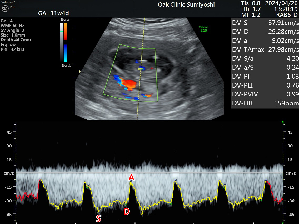
We check the ease of blood flow from the umbilical cord to the baby’s heart (PI). S: Systole, D: Diastole, a: Atrial contraction. PI (Pulsatility Index) is calculated, with a normal value being 1.5 or lower.
If this value is high, it indicates increased vascular resistance, which may raise concerns about fetal hypoxia and potentially affect fetal development.
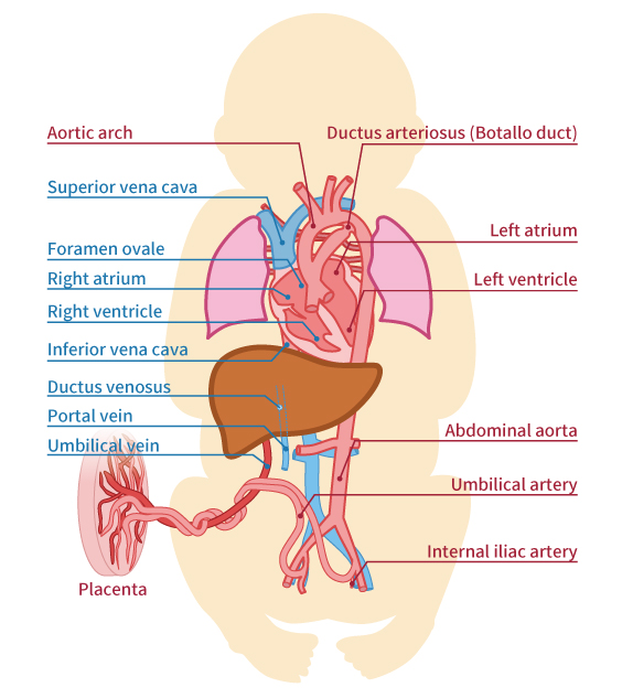
The umbilical cord, which connects the fetus and placenta, is about 50 cm long and 2 cm in diameter by the 10th month of pregnancy. It contains two arteries and one vein. The arteries carry blood from the fetus’s heart to the placenta, while the vein carries blood from the placenta back to the fetus. One of the umbilical arteries is a branch of the internal iliac artery within the fetus’s body, and as shown in the ultrasound image (A), it travels through both sides of the bladder (B) in the fetal pelvis before ascending to the umbilicus.
On the other hand, the umbilical vein inside the fetus ascends from the umbilicus to the liver and ductus venosus. In the case of a single umbilical artery, one of the arteries has merged with the other, resulting in a single artery.
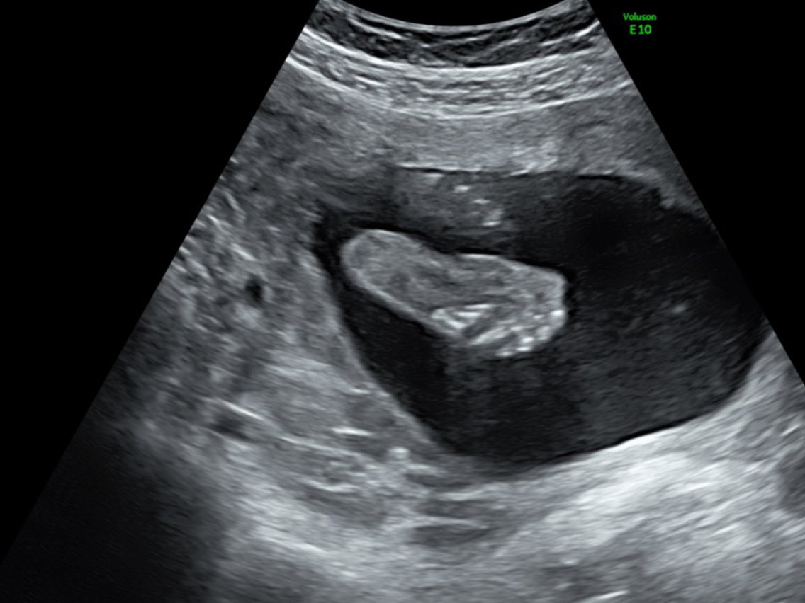
The Length from the Baby’s Head to the Bottom
Between weeks 8 and 11 of pregnancy, there is little individual variation in fetal development, so the length from the baby’s head to the bottom (CRL: Crown-Rump Length) can be used to determine the due date and the number of weeks of pregnancy.
After week 12 of pregnancy, the BPD (Biparietal Diameter: the largest diameter of the baby’s head from side to side) is measured to monitor the baby’s development.

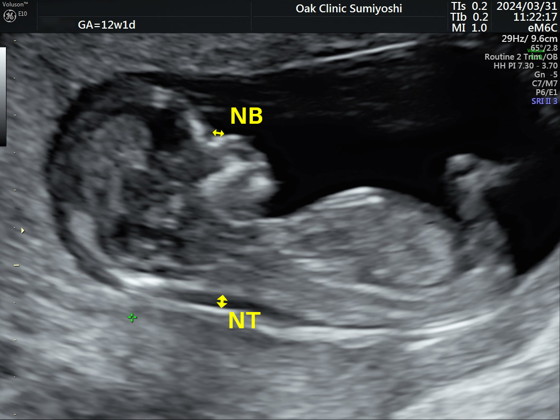
NT (Nuchal Translucency) refers to the swelling at the back of the baby's neck seen around weeks 11 to 13 of pregnancy, and it is present to some degree in all babies. NB (Nasal Bone) refers to the nasal bone. The standard measurements are: NT should be less than 3mm, and NB should be greater than 1.5mm.
An increased NT or a missing nasal bone may raise the possibility of chromosomal abnormalities, but many babies with a NT of 5mm are born without any diseases. It is not accurate to diagnose a condition based solely on the thickness of the NT.
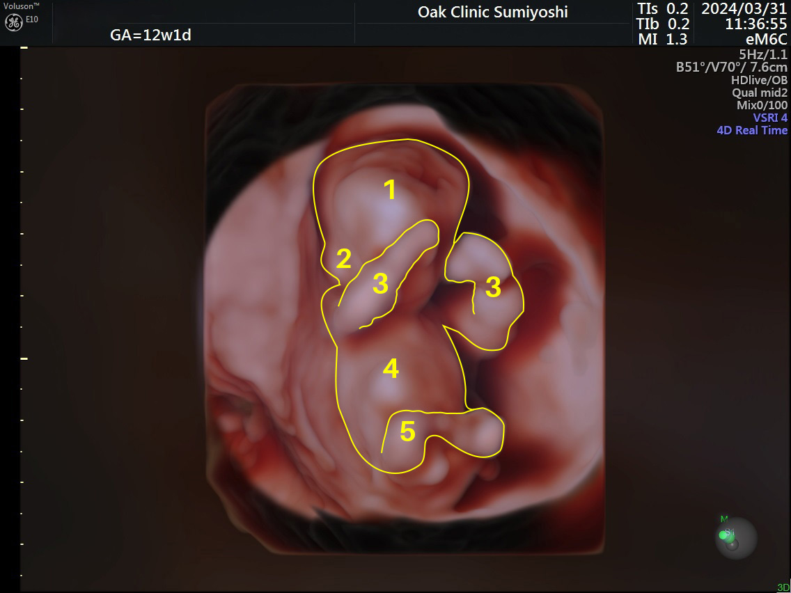
This is the fetus seen using HDlive. 1 is the head, 2 is the ear, 3 are the arms on both sides, 4 is the torso, and 5 is the legs.
Our clinic has introduced 4D and HDlive-equipped ultrasound technology, which has enhanced both the expressive capability and imaging accuracy. By changing the position of the light source, shadows are created, allowing us to provide realistic 3D images.
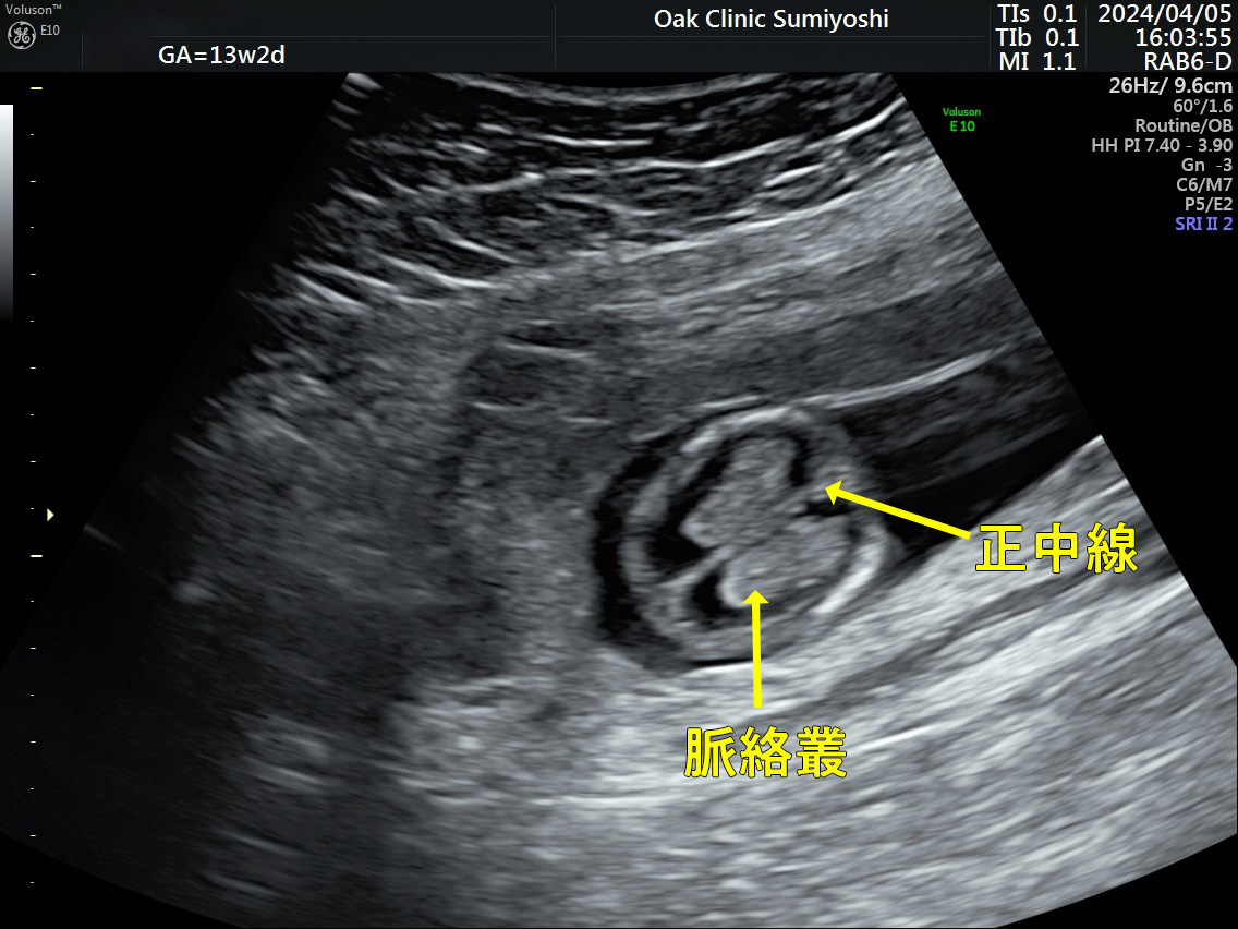
This is a transverse section of the fetal head. We observe whether the structures inside the skull are symmetrical along the midline, whether there are any cysts (sac-like lesions) along the midline, and whether there are any cysts or abnormalities in the size of the choroid plexus (the main organ for producing cerebrospinal fluid) in the brain.
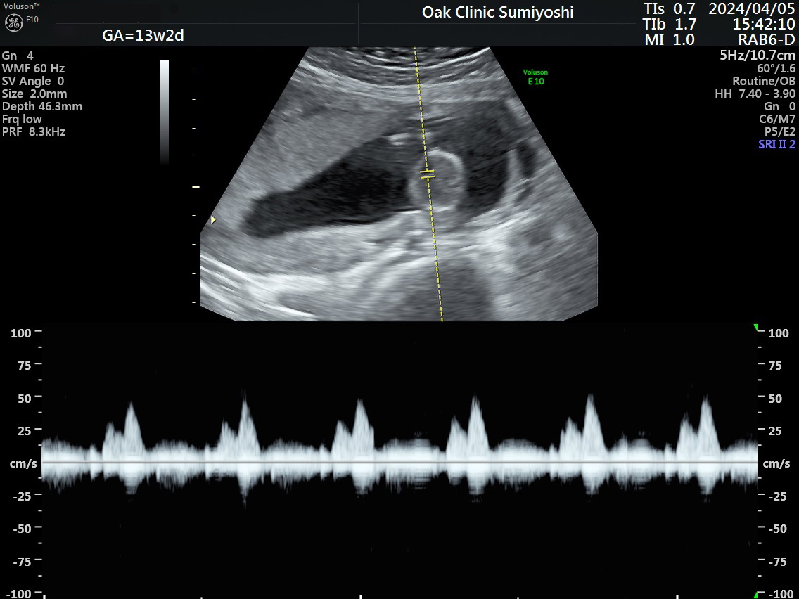
The tricuspid valve is the valve located between the right atrium and right ventricle of the heart, one of the four chambers.
We measure the blood flow from the right atrium to the right ventricle.
In the image, there is no regurgitation, and the waveform is normal. If there is regurgitation, the waveform indicating blood flow below the echo image will be significantly larger than the central line. In some cases, regurgitation can be observed in normal babies as well, but the standard blood flow velocity is below 60 cm/s.
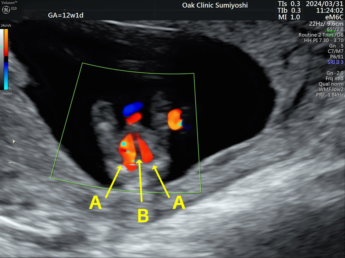
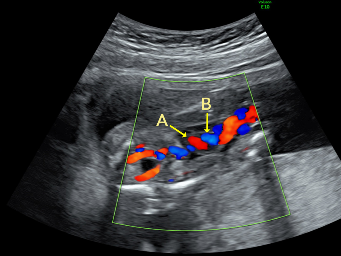
In the color Doppler method, blood flow towards the ultrasound probe is shown in red, and flow away from the probe is shown in blue. The umbilical cord contains two thin arteries (umbilical arteries) wrapped around one thick vein (umbilical vein). The umbilical arteries carry venous blood, while the umbilical vein carries arterial blood.
In the ultrasound image, you can see that the two umbilical arteries are carrying blood towards the placenta (A), and the single umbilical vein is carrying blood from the placenta to the fetus (B).
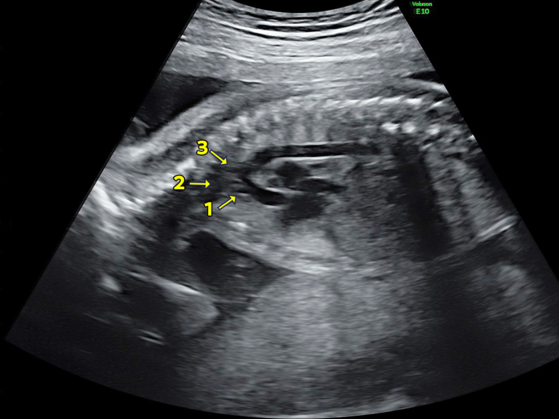
The portion of the aorta that curves in an arch shape as it exits the fetal heart is called the aortic arch. We examine whether there are any narrow areas (stenosis or dissection) in the aortic arch, and check whether it connects properly to the brachiocephalic artery (1), left common carotid artery (2), and left subclavian artery (3), in order to confirm there are no abnormalities in the course of the vessels.
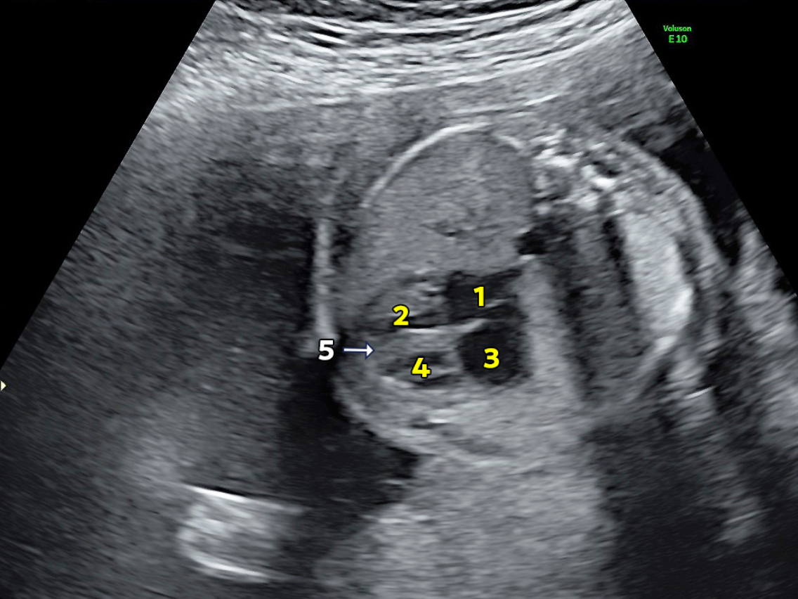
The heart is divided into four chambers: the right atrium, right ventricle, left atrium, and left ventricle. The atria receive blood, while the ventricles pump blood out. Additionally, there are the atrial septum between the atria and the ventricular septum between the ventricles. The atrial septum has a gap (the foramen ovale) during fetal development, which naturally closes after birth. The ventricular septum, which divides the originally single ventricle into two chambers, is formed between the 4th and 8th weeks of pregnancy.
However, if the ventricular septum is incomplete or there is a defect (hole), it is called a ventricular septal defect (VSD). VSD is the most common congenital heart condition, and depending on the location and size of the defect, it may close naturally as the baby grows, or surgery may be required.
This ultrasound image shows the right atrium (1), right ventricle (2), left atrium (3), left ventricle (4), as well as the ventricular septum (5) between the right ventricle (2) and the left ventricle (4).
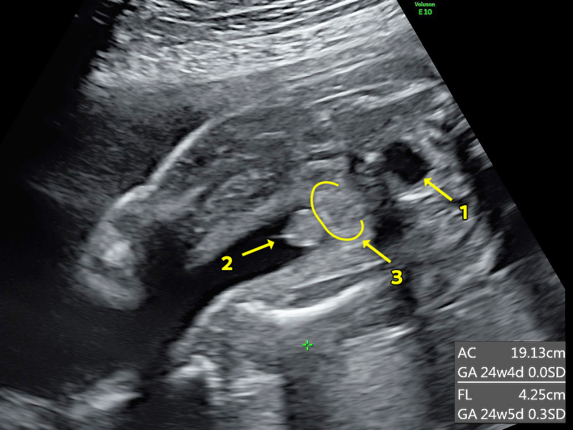
The baby's gender can be identified around 14–15 weeks for boys and 17–18 weeks for girls.
For boys, the penis and scrotum can be seen, while for girls, the area between the legs appears leaf-shaped or coffee bean-shaped.
This ultrasound shows a boy. The protrusion in front of the bladder (1), penis (2), and scrotum (3) are visible.
This is an ultrasound image of the baby’s foot from the side. (1) Sole, (2) Toes, (3) Ankle.
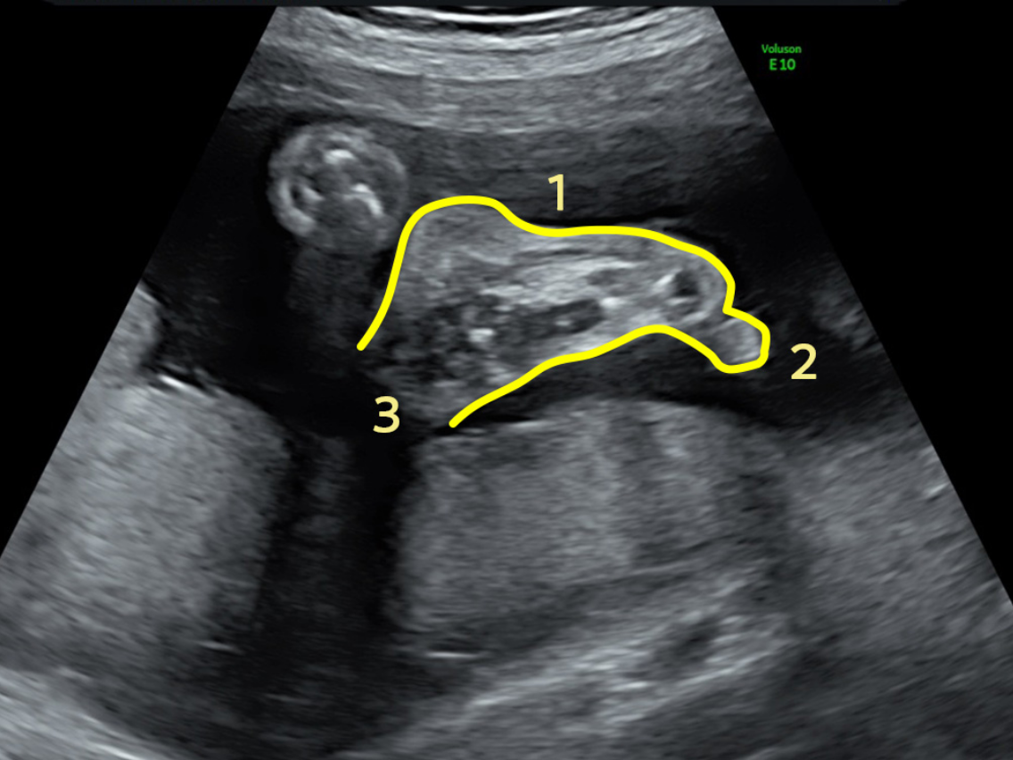
The hands and feet begin to form around weeks 8–9 of pregnancy, and by week 10, the webbed fingers and toes start to separate. The ultrasound checks for arch formation in the soles and the number of toes.
| Q | What is the difference between the ultrasound performed during routine prenatal check-ups and the detailed fetal ultrasound? |
| A | The ultrasound performed during routine prenatal check-ups mainly checks the mother's health, while the detailed fetal ultrasound examines the baby's health. Although fetal size and amniotic fluid levels are also checked during routine prenatal check-ups, the time available for each pregnant woman is limited. To thoroughly examine the baby's health from head to toe, including the placenta and amniotic fluid levels, it is necessary to dedicate time to check specific items according to the appropriate stage of pregnancy. |
| Q | How long does the test take? |
| A | The doctor’s consultation and ultrasound examination are scheduled for a 1-hour slot. At the end of the ultrasound examination, HD-Live/4D imaging is also provided. |
| Q | What does the fetal detailed ultrasound examination check for? |
| A | It checks the entire body, including the fetal heart, brain, face, spine, major blood vessels, reproductive organs, limbs, lungs, stomach, and intestines, as well as the placenta, umbilical cord, and amniotic fluid volume. |
| Q | Is it okay to choose just one stage: early, mid, or late term? |
| A | The items checked differ depending on the stage, so it is recommended to undergo each stage. In the early stage, we check for major abnormalities such as anencephaly, significant heart abnormalities, situs inversus, abdominal wall defects, umbilical hernia, and limb defects. Additionally, neck swelling (NT) is only assessed in the early stage. In the mid and late stages, as the fetus grows, we check for more detailed structural abnormalities that could not be confirmed in the early stage. Specifically, we examine brain structures, blood flow, heart vessel pathways, structural abnormalities, and developmental issues in the urinary and reproductive organs, among other things. The number of items to be checked is doubled compared to the early stage. |
| Q | If the items checked in the mid and late stages are more than in the early stage, is the early stage unnecessary? |
| A | The number of items increases as the fetus grows, but it is considered more beneficial to detect issues at an earlier stage. Even if you and your partner are prepared to accept any potential conditions, discovering them earlier allows you to choose the appropriate fetal treatments, delivery location, and timing. Since routine ultrasound scans during prenatal check-ups are limited, adding early-stage fetal detailed ultrasound scans has significant value. |
English Help Desk
TEL:070-1820-0909
Email:english_help@oakclinic-group.com
[Reception time]
Monday-Saturday 09:00-17:00
For urgent inquiries, please contact us by phone.